he winners of the 2012 Nikon International Small World photography contest were announced Oct. 23. The competition, launched in 1974, honors those spectacularpictures taken through a microscope, called photomicrographs. Each year a panel of judges picks the top 20, plus honorable mentions.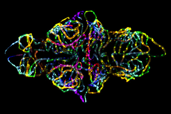 First place went to this photo showing the blood-brain barrier in a live zebrafish embryo. 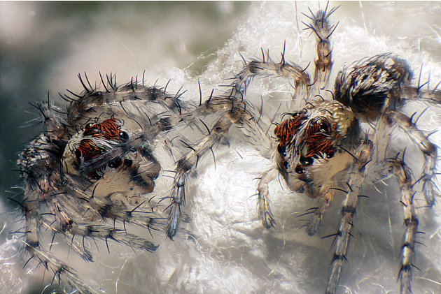 Live newborn lynx spiderlings shot using Reflected Light, Fiber Optics and Image Stacking Techniques at six times magnification. This image received second place. (Walter Piorowski) 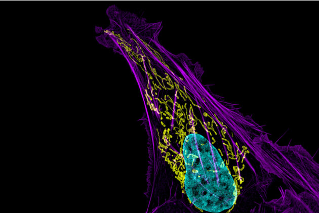 Human bone cancer (osteosarcoma) showing actin filaments (purple), mitochondria (yellow), and DNA (blue) captured with the Structured Illumination Microsopy (SIM) technique at 63 times magnification. This image received third place. (Dr. Dylan Burnette/National Institutes of Health) 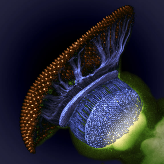 This space-y photo of a fruit fly (Drosophila melanogaster) visual system halfway through pupal development took home fourth prize. Image shows the fruit fly's retina (gold), photoreceptor axons (blue) and brain (green). 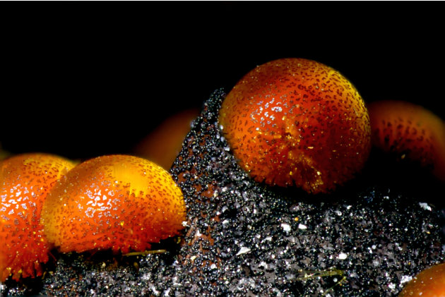 Cacoxenite (mineral) from La Paloma Mine, Spain in the Transmitted Light technique at 18 times magnification. This image received fifth place. (Honorio Cócera-La Parra/University of Valencia Museum of Geology, Department of Geology) 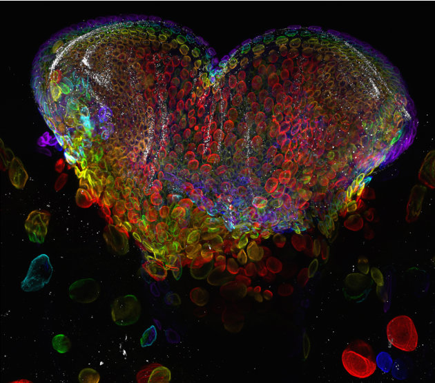 Eye organ of a Drosophila melanogaster (fruit fly) third-instar larvae pictured in the Confocal technique at 60 times magnification. This image received seventh place. (Dr. Michael John Bridge/University of Utah HSC Core Research Facilities - Cell Imaging Lab) 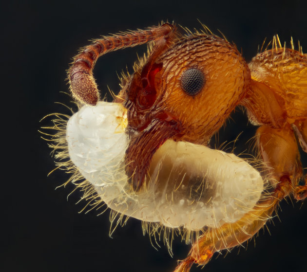 Myrmica sp. (ant) carrying its larva captured using Reflected Light and Image Stacking Techniques at five times magnification. This image received ninth place. (Geir Drange) 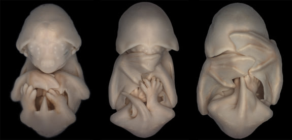 To capture the development of bat embryos of the species Molossus rufus, the black mastiff bat, Dorit Hockman placed the embryos of different ages side-by-side. This image received 20th place. |
Ads
Space For Content and Image Ads
Contact: mohibnabigsm@hotmail.com
Winning photos showcase microscopic world
Posted by
Anonymous
Subscribe to:
Post Comments (Atom)
Post a Comment